JavaScript is disabled for your browser. Some features of this site may not work without it.
We are excited to announce that the repository will soon undergo an upgrade, featuring a new look and feel along with several enhanced features to improve your experience. Please be on the lookout for further updates and announcements regarding the launch date. We appreciate your support and look forward to unveiling the improved platform soon.
| dc.contributor.other | University of Pretoria. Faculty of Veterinary Science. Dept. of Companion Animal Clinical Studies | |
| dc.contributor.upauthor | Van Schoor, Mirinda | |
| dc.date.accessioned | 2010-11-05T06:16:42Z | |
| dc.date.available | 2010-11-05T06:16:42Z | |
| dc.date.created | 2007 | |
| dc.date.issued | 2010-11-05T06:16:42Z | |
| dc.description | Metadata assigned by Dr. M. van Schoor, Senior Lecturer, Dept. of Companion Animal Clinical Studies | en |
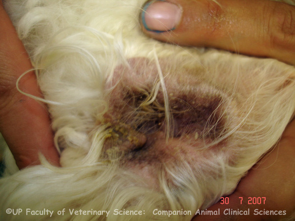
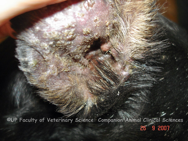
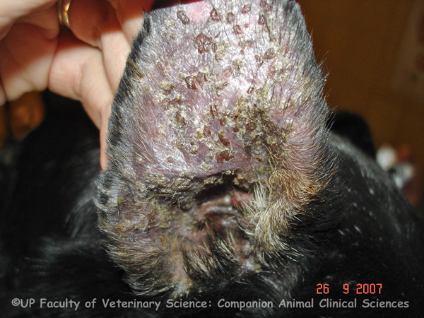
| dc.description.abstract | PHOTOS 1-3: Otitis externa is an acute or chronic inflammatory disease of the external ear predisposed to by increased moisture and decreased ventilation in the ear canal. Symptoms of otitis externa include otic pruritis, head rubbing, ear scratching, head shaking, aural haematomas, erythema and head tilt. A malodorous discharge may be present. In acute cases the inner ear pinna and ear canal are erythematous and swollen. Erythema of the external ear canal and pinna with waxy pale exudates can be caused by an underlying allergy and yeast infection. Erosion and ulceration of the ear canal may also occur. Alopecia of the pinna and crusts are also common. Exudates form crusts. Diagnosis of otitis externa is based on history and clinical findings. Otoscopic examination, microscopy and cytology of ear swabs are used to diagnose otitis and the aetiological agent. Radiography or computed tomography can be used to diagnose bullous involvement. The primary causes of otitis externa include parasites, foreign bodies, hypersensitivities, keratinization disorders, endocrine disorders, autoimmune diseases, inflammatory polyps and neoplasia. The primary cause of otitis can be determined by dermatohistopathology. Secondary causes of otitis include bacterial infection, yeast infection, chronic pathologic changes and otitis media. There are different treatments for the different types of otitis. Treatment options include treating the ears with a drying agent after the dog gets wet, ear cleaning with a ceruminolytic agent and treatment of underlying allergies. Cocker spaniels and long haired dogs are especially prone to otitis externa. Demodicosis can also cause crusting otitis. Pemphigus foliaceus can also cause crusting on the pinnae of the ears. PHOTOS 4-5: Examining the discharge may be useful to determine the primary cause of otitis externa. For example, dry black exudate indicates ear mite or Malassezia infection. A purulent yellow discharge is indicative of a Gram negative bacterial infection, especially Pseudomonas. However, the appearance of the discharge alone is not reliable enough to determine cause and other tests such as culturing or cytology should be used to confirm the diagnosis. PHOTOS 6-8: Malassezia pachydermatis is a yeast that is normally present in small numbers in the external ear canal of dogs. Malassezia pachydermatis commonly causes otitis externa in West Highland White Terriers and Basset Hounds. Long haired breeds and those with pendulous ears are predisposed to otitis externa. The ear canals of these breeds contain more apocrine and sebaceous glandular tissue. In cases of Malassezia otitis the ears become very erythematous, malodorous and hyperplastic. Otitis externa results in increased production of cerumen and increased cerumen gland activity. The cerumen may be thick and oleaginous containing a large amount of yeast. Atopy of hirsute canals may favour colonization by Malassezia pachydermatis. Overgrowth of Malassezia pachydermatis may occur secondary to underlying diseases like diabetes mellitus or due to an internal malignancy. | en |
| dc.description.abstract | REFERENCES: 1. Foster, A & Foil, C (eds) 2003, 'BSAVA manual of small animal dermatology', 2nd ed., British Small Animal Veterinary Association, Gloucester, pp. 104-111. 2. Harvey, RG, Harari, J & Delauche, AJ 2001, ‘Ear diseases of the dog and cat’, Manson Publishing, London, pp.114-116. 3. Mactaggart, D 2008, 'Assessment and management of chronic ear disease', In Practice, vol. 30, no. 8, pp. 450-458. [http://0-inpractice.bvapublications.com]. 4. Medleau, L & Hnilica, KA 2006, ‘Small Animal Dermatology: A Color Atlas and Therapeutic Guide’, 2nd ed., Saunders Elsevier, St. Louis, pp. 64-65, 190-191, 376-388. 5. Patterson, S 1998, 'Skin Diseases of the Dog', Blackwell Sciences, Oxford, pp. 139-152. 6.Scott, DW, Miller, WH, Griffin, CE 2001, 'Muller & Kirk’s small animal dermatology', 6th ed., W.B. Saunders Company, Philadelphia, p. 1204. | en |
| dc.format.extent | 8 colour photos | en |
| dc.format.medium | JPEG | en |
| dc.identifier.uri | http://hdl.handle.net/2263/15193 | |
| dc.relation.ispartofseries | Veterinary critical care slide collection (Dr M. van Schoor) | en |
| dc.rights | © Dr Mirinda van Schoor, University of Pretoria. Dept. of Companion Animal Clinical Studies (Original and digital). Provided for educational purposes only. It may not be downloaded, reproduced or distributed in any format without written permission of the original copyright holder. Any attempt to circumvent the access controls placed on this file is a violation of copyright laws and is subject to criminal prosecution. Please contact the collection administrator for copyright issues. | en |
| dc.subject | Veterinary intensive care | en |
| dc.subject | Basset hound | en |
| dc.subject | Crusting | en |
| dc.subject | Ear | en |
| dc.subject | Erythema | en |
| dc.subject | Demodex | en |
| dc.subject | Malassezia otitis | en |
| dc.subject | Malassezia pachydermatis | en |
| dc.subject | Malodorous discharge | en |
| dc.subject | Pemphigus foliaceus | en |
| dc.subject | West highland white terrier | en |
| dc.subject.lcsh | Veterinary critical care | en |
| dc.subject.lcsh | Veterinary medicine -- South Africa | en |
| dc.subject.lcsh | Veterinary emergencies | en |
| dc.title | Otitis externa | en |
| dc.type | Still Image | en |