JavaScript is disabled for your browser. Some features of this site may not work without it.
| dc.contributor.other | University of Pretoria. Faculty of Veterinary Science. Dept. of Companion Animal Clinical Studies | |
| dc.contributor.upauthor | Van Schoor, Mirinda | |
| dc.date.accessioned | 2010-11-05T06:40:58Z | |
| dc.date.available | 2010-11-05T06:40:58Z | |
| dc.date.created | 2008 | |
| dc.date.issued | 2010-11-05T06:40:58Z | |
| dc.description | Metadata assigned by Dr. M. van Schoor, Senior Lecturer, Dept. of Companion Animal Clinical Studies | en |
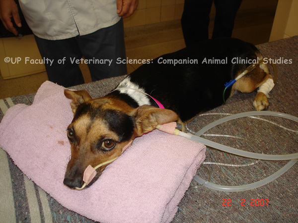
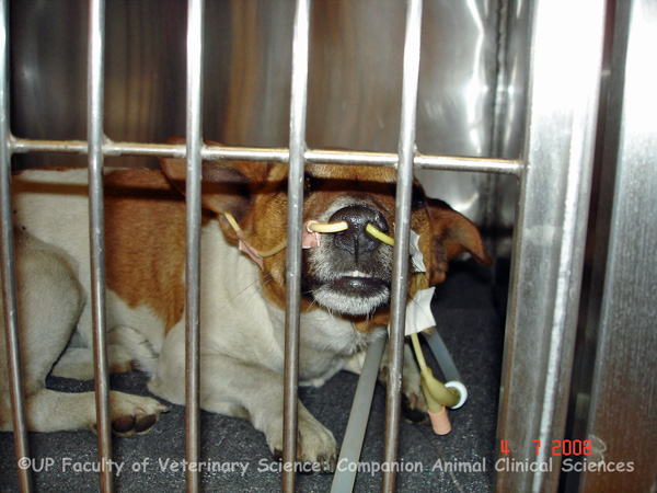
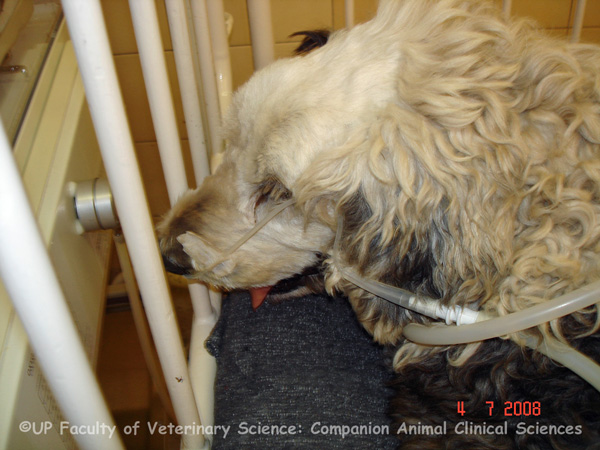
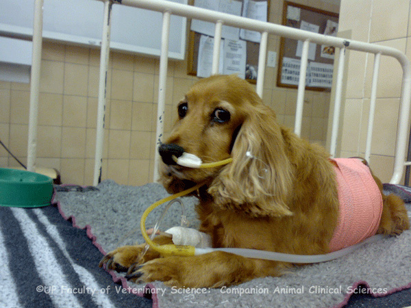
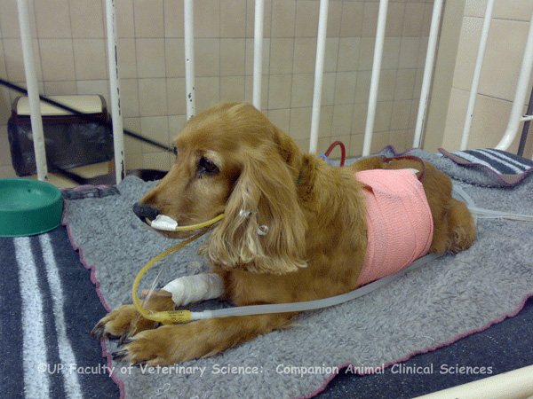
| dc.description.abstract | PHOTOS 1-5: Many critical illnesses can cause hypoxia to occur and oxygen supplementation may be necessary to improve oxygen delivery and to prevent lactic acidosis. A nasal cannula is the best way to supplement oxygen in conscious patients. Oxygen supplementation is an important management tool for critical conditions such as cardiopulmonary disease, sepsis, SIRS and head trauma. Nasal cannulas are easy to place, require minimal equipment and are well tolerated by patients. To place a nasal cannula, the patient's nasal passage is anaesthetized and the correct length of tube is measured from the nostril to the medial canthus of the eye. The lubricated tube is gently inserted into the ventral nasal meatus and is secured adjacent to the nostril with suture or staples. Sneezing and intolerance can be alleviated by reapplying the topical anaesthetic or pushing the nasal cannula into the nasopharyngeal region. An Elizabethan collar can be used to prevent the patient from dislodging the nasal cannula. A nasal cannula allows for prolonged oxygen delivery and permits access to the patient for examination and treatment purposes without losing the oxygen rich environment. A disadvantage of using a nasal cannula is that the fraction of inspired oxygen cannot always be determined accurately. The necessary oxygen flow rate is based on patient size, respiratory rate, respiratory pattern and the degree of open-mouth breathing. If a unilateral nasal cannula is used it should be replaced with a new cannula on the opposite side every 48 hours so as to reduce damage to the airway. Most animals tolerate the cannula well. | en |
| dc.description.abstract | REFERENCES: PHOTOS 1-5: 1. Manning, AM 2002, ‘Oxygen therapy and toxicity’, Veterinary Clinics of North America. Small Animal Practice, vol. 32, no. 5, pp. 1005-1020. 2. Silverstein, DC & Hopper, K (eds) 2009, ‘Small animal critical care medicine’, Saunders Elsevier, St. Louis, pp. 78-80. | en |
| dc.format.extent | 5 colour photos | en |
| dc.format.medium | JPEG | en |
| dc.identifier.uri | http://hdl.handle.net/2263/15207 | |
| dc.relation.ispartofseries | Veterinary critical care slide collection (Dr M. van Schoor) | en |
| dc.rights | © Dr Mirinda van Schoor, University of Pretoria. Dept. of Companion Animal Clinical Studies (Original and digital). Provided for educational purposes only. It may not be downloaded, reproduced or distributed in any format without written permission of the original copyright holder. Any attempt to circumvent the access controls placed on this file is a violation of copyright laws and is subject to criminal prosecution. Please contact the collection administrator for copyright issues. | en |
| dc.subject | Veterinary intensive care | en |
| dc.subject | Hypoxia | en |
| dc.subject | Cardiopulmonary diseases | en |
| dc.subject | Sepsis | en |
| dc.subject | Head trauma | en |
| dc.subject | Elizabethan collar | en |
| dc.subject | Respiration | en |
| dc.subject | Nasal cannula | en |
| dc.subject.lcsh | Veterinary critical care | en |
| dc.subject.lcsh | Veterinary medicine -- South Africa | en |
| dc.subject.lcsh | Veterinary emergencies | en |
| dc.title | Oxygen therapy | en |
| dc.type | Still Image | en |