JavaScript is disabled for your browser. Some features of this site may not work without it.
| dc.contributor.other | University of Pretoria. Faculty of Veterinary Science. Dept. of Companion Animal Clinical Studies | |
| dc.contributor.upauthor | Van Schoor, Mirinda | |
| dc.date.accessioned | 2010-11-05T06:04:24Z | |
| dc.date.available | 2010-11-05T06:04:24Z | |
| dc.date.created | 2008 | |
| dc.date.issued | 2010-11-05T06:04:24Z | |
| dc.description | Metadata assigned by Dr. M. van Schoor, Senior Lecturer, Dept. of Companion Animal Clinical Studies | en |
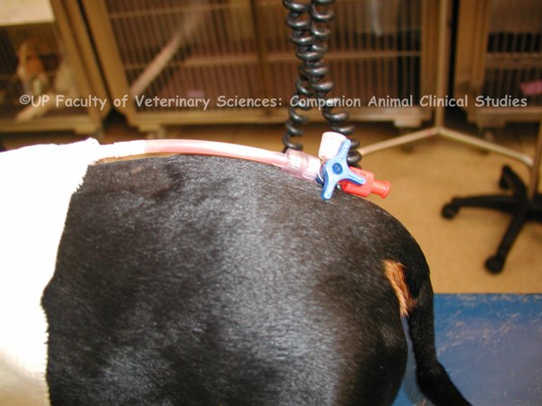
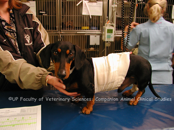
| dc.description.abstract | PHOTO 1: Chylothorax is the accumulation of chyle from the thoracic duct in the thoracic cavity. The fluid contains triglycerides, lymphocytes, protein and fat soluble vitamins. The accumulation of chyle is due to abnormal pressure in the thoracic duct which leads to exudation of chyle from intact but dilated thoracic lymph vessels. The chylous effusion causes fibrosing pleuritis which interferes with the ability of the lungs to expand. On physical examination the heart and lung sounds are muffled, there are increased bronchovesicular sounds; cyanosis and pale mucous membranes can be seen. Traumatic chylothorax may occur if trauma causes the thoracic duct to rupture. Non traumatic chylothorax may be caused by generalized lymphangiectasis, inflammation or obstruction of lymphatic flow by neoplasia or increased venous pressure. Other causes of non traumatic chylothorax include mediastinal lymphoma in cats, cardiomyopathy, pericardial disease, right heart failure, lung lobe torsion and diaphragmatic hernia. Congenital chylothorax predisposes to chylothorax later in life. Traumatic events like surgery or being hit by a car can cause chylothorax in these animals. Clinical signs of chylothorax include respiratory distress, lethargy, anorexia, weight loss and exercise intolerance. Thoracocentesis and fluid therapy are used to stabilise animals with chylothorax. PHOTOS 2-3: A chest drain (thoracocentesis) may be necessary to stabilize an animal suffering from chylothorax. Chest drains may be required every 1 to 2 weeks in the initial stages of treatment. The length of intervals of thoracocentesis will depend on the owners’ observation of when the animal displays signs of increased respiratory effort or decreased activity. If the patient responds well to medical management the intervals of thoracocentesis can be gradually decreased. PHOTOS 4-9: Pericardial tap or pericardiocentesis is the use of a catheter to remove a volume of pericardial effusion for diagnostic and therapeutic purposes. It is used when pericardial effusion causes cardiac tamponade and when a pericardial effusion of unknown aetiology is present. It may also be necessary to remove chyle from the pleural cavity in animals suffering from chylothorax. Ultrasound guidance prior to the procedure to locate an area of maximal pericardial effusion and minimal lung is useful. The animal is placed in left lateral recumbency and ultrasound is used to confirm that pericardial effusion is present in sufficient volume for centesis. The area is widely clipped and aseptically prepared. The site for centesis is again confirmed via ultrasound. The area is infused subcutaneously, intramuscularly and subpleurally with Lidocaine. A syringe-catheter-stylet combination is advanced while drawing back on the syringe plunger to create negative pressure. A flashback of effusion is seen in the syringe and the needle is advanced 2-3 mm further into the chest. The catheter is advanced a few centimetres further into the pericardial space. The effusion is withdrawn using a large volume syringe until the pericardium is empty. Once the effusion has been removed the catheter is withdrawn. The skin incision may be closed using tissue glue. | en |
| dc.description.abstract | REFERENCES: PHOTOS 1-9: 1. Côte, E (ed) 2007, ‘Clinical veterinary advisor : dogs and cats’, Mosby Elsevier, St. Louis, pp. 1298-1299. 2. Nelson, RW & Couto, CG (eds) 2009, ‘Small animal internal medicine’, 4th ed., Mosby Elsevier, St. Louis, pp.338-339. 3. Tilley, LP & Smith, FWK 2004, ‘The 5-minute veterinary consult : canine and feline’, 3rd ed., Lippincott Williams & Wilkins, Baltimore, pp. 230-231. | en |
| dc.format.extent | 9 colour photos | en |
| dc.format.medium | JPEG | en |
| dc.identifier.uri | http://hdl.handle.net/2263/15181 | |
| dc.relation.ispartofseries | Veterinary critical care slide collection (Dr M. van Schoor) | en |
| dc.rights | © Dr Mirinda van Schoor, University of Pretoria. Dept. of Companion Animal Clinical Studies (Original and digital). Provided for educational purposes only. It may not be downloaded, reproduced or distributed in any format without written permission of the original copyright holder. Any attempt to circumvent the access controls placed on this file is a violation of copyright laws and is subject to criminal prosecution. Please contact the collection administrator for copyright issues. | en |
| dc.subject | Veterinary intensive care | en |
| dc.subject | Cardiac tamponade | en |
| dc.subject | Catheter | en |
| dc.subject | Chest drain | en |
| dc.subject | Fluid therapy | en |
| dc.subject | Pericardial catheter | en |
| dc.subject | Pericardial effusion | en |
| dc.subject | Pericardial tap | en |
| dc.subject | Pericardiocentesis | en |
| dc.subject | Pleural cavity | en |
| dc.subject | Pleuritis | en |
| dc.subject | Respiratory distress | en |
| dc.subject | Thoracic duct | en |
| dc.subject | Thoracocentesis | en |
| dc.subject | Trauma | en |
| dc.subject | Ultrasound | en |
| dc.subject.lcsh | Veterinary critical care | en |
| dc.subject.lcsh | Veterinary medicine -- South Africa | en |
| dc.subject.lcsh | Veterinary emergencies | en |
| dc.title | Chyle accumulation due to chylothorax | en |
| dc.type | Still Image | en |