JavaScript is disabled for your browser. Some features of this site may not work without it.
| dc.contributor.other | University of Pretoria. Faculty of Veterinary Science. Dept. of Companion Animal Clinical Studies | |
| dc.contributor.upauthor | Van Schoor, Mirinda | |
| dc.date.accessioned | 2010-11-03T08:23:56Z | |
| dc.date.available | 2010-11-03T08:23:56Z | |
| dc.date.created | 2007 | |
| dc.date.issued | 2010-11-03T08:23:56Z | |
| dc.description | Metadata assigned by Dr. M. van Schoor, Senior Lecturer, Dept. of Companion Animal Clinical Studies | en |
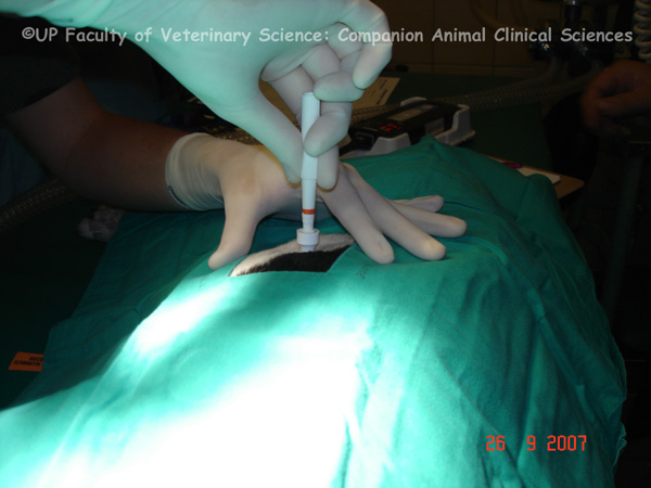
| dc.description.abstract | PHOTO 1: A preoperative biopsy should be done if it will help choose a therapeutic modality and the degree of therapy. The equipment needed for a biopsy include a biopsy punch, a scalpel blade, local anaesthetic such as Lidocaine, a sterile surgical drape, surgical scrub, sterile surgical gloves and suture materials. Biopsy punches are disposable and range from 2 to 6mm in diameter. PHOTOS 2-3: Larger biopsies are usually preferred by the pathologist for histological diagnosis. The junction between normal and abnormal tissue should be biopsied. Punch biopsies are used to get skin samples. The skin directly over the selected site is clipped and cleaned with a surgical scrub. A local anaesthetic is injected around the lesion and the biopsy site is scrubbed again. The skin is stretched and the biopsy punch is placed at right angles to the skin surface. The punch is rotated in one direction and firm downward pressure is applied. The punch is then angled parallel to the skin while still applying pressure. The punch is rotated to sever the base of the biopsied material and is then removed. The tissue core is elevated with the point of a needle and the base severed with a scalpel. The incision is sutured closed. | en |
| dc.description.abstract | REFERENCES: PHOTOS 1-3: 1. Hahn, KA 2002, ‘Veterinary oncology’, Butterworth-Heinemann, Boston, pp.50. 2. McCurnin, DM & Poffenbarger, EM 1991, ‘Small animal physical diagnosis and clinical procedures’, W.B. Saunders , Philadelphia, pp.192-193. | en |
| dc.format.extent | 3 colour photos | en |
| dc.format.medium | JPEG | en |
| dc.identifier.uri | http://hdl.handle.net/2263/15145 | |
| dc.relation.ispartofseries | Veterinary critical care slide collection (Dr M. van Schoor) | en |
| dc.rights | © Dr Mirinda van Schoor, University of Pretoria. Dept. of Companion Animal Clinical Studies (Original and digital). Provided for educational purposes only. It may not be downloaded, reproduced or distributed in any format without written permission of the original copyright holder. Any attempt to circumvent the access controls placed on this file is a violation of copyright laws and is subject to criminal prosecution. Please contact the collection administrator for copyright issues. | en |
| dc.subject | Veterinary intensive care | en |
| dc.subject | Skin biopsy | en |
| dc.subject | Biopsy set | en |
| dc.subject | Biopsy punch | en |
| dc.subject.lcsh | Veterinary critical care | en |
| dc.subject.lcsh | Veterinary medicine -- South Africa | en |
| dc.subject.lcsh | Veterinary emergencies | en |
| dc.title | Skin biopsy | en |
| dc.type | Still Image | en |